Small animal veterinary case study – Betty Bell the spaniel
History
Betty was presented on 14/5/14. During an echo to investigate a murmur, it had been noted that she had cranial abdominal pain so she was referred for investigation and an abdominal scan. Appetite was good and body weight was stable. She was not PUPD.
Physical examination
On clinical examination, she was bright and alert and in good body condition at 15.5kg. There was evidence of cranial abdominal pain. A grade IV/VI systolic murmur was auscultated. Physical exam was otherwise unremarkable.
Laboratory
In house clotting times (PT and APTT) were within reference interval. Platelet count was adequate for haemostasis and PCV was 37%. Venous blood gases were unremarkable. Haematology and biochemistry were fairly unremarkable
Diagnostic imaging
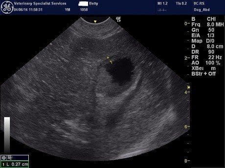
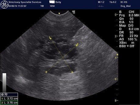
An abdominal ultrasound was performed. There was no free fluid and no enlarged abdominal lymph nodes. Both kidneys showed increased cortical echogenicity which is a common non-specific age-related change. The spleen was unremarkable. The urinary bladder was unremarkable. There was a cystic lesion within the tip of one of the liver lobes on the left side. FNAs were obtained from this area and a dark brown/red fluid was retrieved which had a PCV of 4%. Fluid analysis revealed a cystic fluid containing red blood cells. Some sludge was identified within the gall bladder.
Chest radiographs were unremarkable.
Diagnosis: Cystic liver lesion
Follow-up
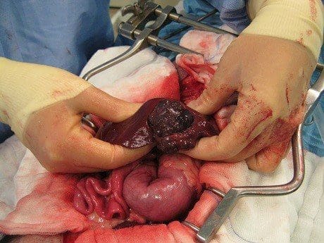
Betty underwent exploratory surgery to remove the affected piece of liver. This was successfully excised and histology confirmed that this was a benign lesion consistent with cystic hyperplasia of the bile ducts. Betty has had an uneventful recovery from surgery