Small animal cardiac ultrasound measurements
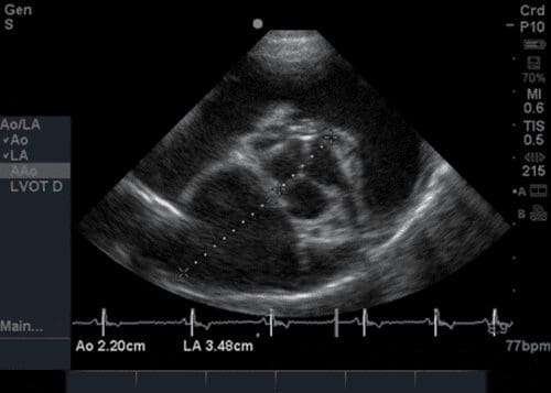
LA:Ao (Left atrium:aortic root ratio)
Right parasternal short axis view at the level of left atrium and aorta (‘Mercedes Benz’)
Measurement on frozen B-mode image
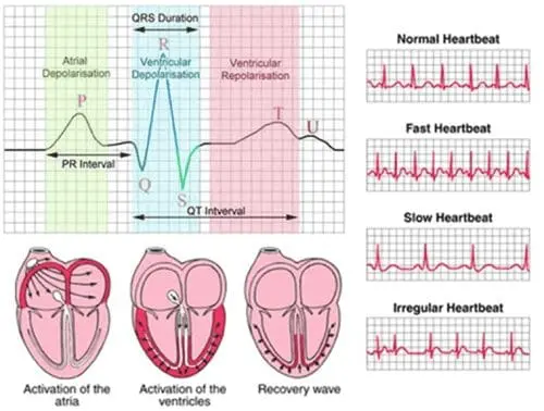
Measurement taken during diastole (just before QRS on ECG)
Measurements taken: LA Diam + Ao Diam (machine will calculate LA:Ao for you)
Lines should be drawn through middle of aortic valve leaflets and continued through left atrium in a straight line
Normal value – <1.5 in normal dog
Increased value in left atrial enlargement (disease like Mitral regurgitation)
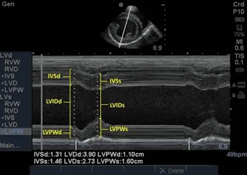
FS (Fractional shortening)
Right parasternal short axis view at level of chordae tendinae (Mushroom view)
Measurement on frozen M-mode image
3 measurements taken during diastole and 3 during systole (6 total)
Diastole (just before QRS on ECG)
– IVSd (Interventricular septum at end diastole)
– LVIDd (Left ventricular internal diameter at end diastole)
– LVPWd (Left ventricular posterior wall thickness at end diastole) or LVFWd
(Left ventricular free wall thickness at end diastole) – depends on if human machine Systole (at nadir of septal motion – when ventricular diameter narrowest)
– IVSs (Interventricular septum at end systole)
– LVIDs (Left ventricular internal diameter at end systole)
– LVPWs (Left ventricular posterior wall thickness at end systole) or LVFWs
(Left ventricular free wall thickness at end systole) – depends on if human machine
All measurements are taken from leading edge to leading edge (see below)
Machine will calculate Fractional shortening
Normal value – >25% in normal dog (breed variations!)
Decreased in DCM (heart contracting poorly during systole)
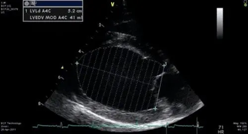
EF (Ejection fraction)
Right parasternal long axis view (4 chamber view)
Measurement on frozen B-mode image
Simpson’s rule used to evaluate left ventricular volume
2 measurements made during diastole and 2 measurements taken during systole
Diastole
– LVLd (Left ventricle length during diastole)
– LVAd (Left ventricle area during diastole)
Systole
– LVLs (Left ventricle length during systole)
– LVAs (Left ventricle area during systole)
Machine will calculate Ejection fraction (will also display EDV and ESV – End diastolic
volume and End systolic volume)
Normal value – >50% in normal dog
Decreased in DCM (heart contracting poorly during systole)